- Ultrasound = sound waves with frequencies > 20,000 Hz (generally > 20 MHz for medical applications)
- Average speed of sound is 1540 m/s. Travels faster through dense material (bone) and slower through air (lung)
- Sound waves travels from probe, through tissue, and reflects back to the probe.
- Sound waves generate voltage through piezoelectric crystals. Gel minimizes impedance difference between tissue and crystal to better transmit waves (can also submerge in saline/water if appropriate).
- The sound wave amplitude is attenuated (reduced) as a function of the distance traveled and the substance attenuation coefficient. Blood most efficient, Lung least efficient. Lower frequency sound is less attenuated
- Attenuation (dB) ~ Frequency (MHz) * depth (cm) / 2
- dB is a log scale. So a 5 MHz frequency at 10 cm depth is attenuated 25 dB, whereas a 10 MHz frequency is attenuated 50 dB. Converting dB to ratio is 10^(dB/10) = 10^(25/10) -> 5 Mhz is ~ 300 fold less attenuated than a 10 MHz frequency. This means that a 5 MHz wave will measure more depth than a higher frequency wave.
- Sector image - one crystal that oscillates/rotates to piece together an image
- Phased transducer - multiple crystals that can operate indepently to be 'phased'
- Linear
- Curvilinear
- Sector looks like curvilinear
- Resolution
- Axial - dependent on frequency/attenuation
- Lateral - dependent on beam width/focusing in a phased transducer
- Temporal - dependent on frame rate
- Hypoechoic/anechoic = black = fluid
- Fluid doesn't stop waves so should see white deep to fluid
- Hyperechoic = white = calcium, fat, gas or "echogenic" tissues
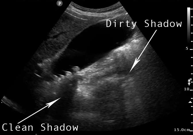
- Clean shadowing (black area behind a white area) = calcium (sound absorbing)
- Dirty shadowing = gas (sound reflecting)
- Refraction = metal
- Probe frequencies
- Low frequency (3-5 Hz) - poor resolution but good penetration (abdominal/renal imaging)
- High frequency (15-18 Hz) - better resolution but lower penetration (e.g. thyroid imaging)
- Dot on side of probe points to patient's right/head (will correlate to screen left. So if you are facing patient, left is left and right is right)
- How to get better pictures: adjust depth, adjust gain, more pressure on probe, more gel
| Body part | Probe | Positioning | Comments |
| Abdomen | |||
| Cardiac/echo | Left lateral decubitus | ||
- B-mode (brightness) = normal 2D ultrasound
- M-mode (motion) = maps motion along a line over time
- Doppler mode = uses Doppler effect to measure blood flow velocity
- Color doppler - color-coded velocity information overlaid on B-mode image
- Continuous wave - Doppler info sampled along a line, and velocity plotted against time
- Pulsed wave - Doppler info sampled from a small 2D sample volume
Artifacts
| Reverberation | ||
| Comet tail | Produced by dense/metallic material, or in lungs at visceral/parietal interface with ARDS/edema | |
| Ring down | Variant of reverberation - 2/2 impedance mismatch generally by gas in lipid/tissue. Air/tissue interface creates impedance mismatch -> reflects sound wave and creates reverberation/ring down artifact. | |
| Mirroring | Reverberation - two strong reflectors in close proximity (e.g. diaphraghm and liver mass). Local reverberations cause delay in return to probe, causing a 'mass' to appear on the other side of the reflector/'mirrored image' | |
| Shadowing | High attenuation reduces transmission past the mass of high attenuation = diminished signal/hypoechoic past the mass | |
| Enhancement | Opposite of shadowing. Low attenuation (e.g. liver cyst) in front of a reflector, so reflected signal is higher and will have brighter area posterior to the cyst. | |